Good time management is crucial in advanced dentistry and new technology is going a long way to help clinicians meet ever demanding schedules. Kent dentist Dr Pedja Pavlovic, a specialist in periodontics and a dental implantologist, uses sophisticated digital imaging to plan and prepare his treatments most effectively and efficiently.
‘Everyone is talking about digital dentistry and how you can use digital imaging systems to provide treatment; from diagnostic procedures and treatment planning to surgical execution, and even the fabrication of the restoration by computers.’
Dr Pavlovic, of the Centre for Aesthetic Periodontics and Implantology in Tunbridge Wells, describes 3D imaging as the first step. ‘Without it you can’t do guided surgery,’ he explains. ‘3D imaging allows you to produce guided templates where you can execute the surgery very efficiently. This reduces clinical time and increases precision.’
AT THE BEGINNING
Dr Pavlovic started his postgraduate training in periodontology at the University of Belgrade, Serbia and completed it in London. He runs a full-time referral practice restricted to periodontics and implant dentistry. He also works in a specialist dental clinic in Harley Street, London.
In Harley Street he uses a different 3D imaging system, but he was so impressed with the Vatech PaX-i3D Green CT that he decided to purchase one for his Kent practice to replace his existing digital imaging system.
‘We are in Tunbridge Wells in the middle of Kent. There aren’t many CT scanners in the area so I would refer patients to London. That was not really a practical proposition – it was inconvenient and time consuming. The ideal scenario was to have an up-to-date imaging system on site that not only provided us with 2D imaging but 3D as well.’
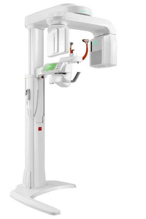
The PaX-i3D Green fitted the bill perfectly. It advances patient safety with lower dose X-rays and a 5.9 second scan time. It also provides four different field of view sizes for CBCT images (5cmx5cm, 8cmx5cm, 8cmx8cm and 12cmx9cm) enabling the clinician to choose the optimum size required while minimising radiation exposure.
Dr Pavlovic visited the Vatech showroom and declared: ‘I need a scanner next week.’ Despite the usual lead times being around five to six weeks, the company said the scanner could be arranged within his timeframe.
‘I was very impressed – they were so efficient. My clinical time is valuable and I didn’t want to spend time with engineers and sales people.’
SETTING UP
A training session was provided to help familiarise Dr Pavlovic and the other clinicians in the practice with the new technology, although he says the new machine has proven ‘very intuitive’ to use. Digital dentistry allows for greater clinical planning and accuracy and the benefits have been immediate.
‘I use the system for analysing pathology around roots for patients needing periodontal treatment and for assessing the anatomy to check the suitability of certain sites for implant placements,’ he says. ‘It goes without saying that careful assessment of patients’ needs and full justification for CBCT imaging is always undertaken.
‘If a CBCT is justified, for which there are very strict guidelines, then instead of opening the flap to do the surgery and encountering unforeseen problems, the surgeon can plan every single detail in front of the computer before the surgery begins. The procedure can then be executed very swiftly and precisely, saving clinical time and benefitting the patient. On a number of occasions you actually detect anatomical details or pathology that you didn’t expect to see there.
‘When everything is prepared, you can go for the implant treatment without any hesitation as you know exactly what to expect.’
Patients respond favourably to the new technology as they appreciate the fact that the dentist can communicate more clearly why treatment is necessary when armed with all the facts.
‘The system also provides the opportunity to combine photographs, radiographs and CBCT on one screen,’ explains Dr Pavlovic. ‘I can load the patient’s clinical photographs and X-rays at the same time on the same screen, which makes the presentation for the patient very smooth… I think it gives patients the confidence that everything is under control. When you present the information with all the images, it’s very reassuring.’
IMPROVING PREDITABILITY
Above all else, Dr Pavlovic says the new technology has increased the ‘predictability of the surgical procedures and the treatments’ carried out at his practice.
‘It is important to have predictable treatment outcomes; I am not just using technology because it’s new. This technology is well proven and in some cases I can’t see that anyone could predictably do the treatment without having this type of imaging.’
While not every practice needs to have a 3D system on site, Dr Pavlovic argues that all patients should have access to this investigation if it is justified and will be beneficial. The PaX-i3D advances patient safety with lower dose X-rays and a 5.9 second scan time. It provides four different field of view sizes for CBCT images, enabling the clinician to choose the optimum size required while minimising radiation exposure.
| PUBLISHED IN: | Implant Dentistry Today, May 2015 |
| DENTIST: | Dr. Pedja Pavlovic |
| EQUIPMENT: | PaX-i3D Green (10x8cm SP) |
| DENTAL PRACTICE: | Centre for Aesthetic Periodontics & Implantology |
| ADDRESS: | 8 Lonsdale Gardens, Tunbridge Wells, Kent, TN1 1NU |
| TELEPHONE: | 01892 617 467 |
| EMAIL: | reception@capiuk.co.uk |
| WEBSITE: | periodonticsandimplants.co.uk |