
‘In the past, dentistry had a poor reputation when it came to patient education and clinician-patient communication,’ says Louise of Hospital Lane Dental and Implant Clinic, Blue Bell Hill.
‘Patients came in and had their treatment and didn’t always know what was going on.’
But at Hospital Lane, a state-of-the-art private clinic that boasts seven surgeries, 11 dentists and a designated imaging room, things couldn’t be more different. Louise, a dentist of 25 years standing, helps guide patients through the initial diagnosis and planning of their treatment, using three-dimensional imagery to show them where a problem lies and how it could be treated.
‘Computing is very much part of dentistry but we are still dealing with human beings. The digital imaging aspect is a real eye-opener for patients. When we show patients the three-dimensional images of their problem areas they can clearly see that we are trying to advise and guide them to the correct treatment options to improve their dental health.
This has the added benefit of improving patient awareness of their dental health and, in turn, their own responsibility for it. As a team we can then work together towards better dental health and wellbeing.’
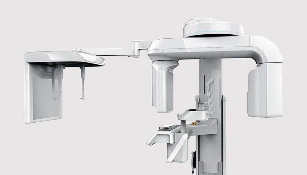
Thanks to its cutting-edge technology, the Hospital Lane clinic is already ahead of the game. The practice uses Vatech PaX-i3D 12×9 SC – an advanced digital dental diagnostic system that incorporates panoramic, cephalometric and three- dimensional CT imaging capabilities in a single machine.
Advanced sensors produce high quality images in two-dimensions and three- dimensions of the dental/maxillofacial regions for dental treatment planning and diagnosis. The PaX-i 3D 12×9 SC provides four different field of volume sizes for CBCT images (5x5cm, 8x5cm, 8x8cm and 12x9cm), enabling the clinician to select the optimum size required while minimising radiation exposur
EARLY INVESTMENTS
In 2001, the practice invested in an early three-dimensional imaging system using tomograms (single slices of X-ray) rather than computerised tomography (layers of X-ray), which had to be developed onto film, traced and carefully interpreted.
However, in 2008, after researching all the imaging machines on offer, the practice switched to the Vatech & E-Woo Technology Picasso Trio SC – the flagship system of its time.
‘The system was digital. We could view the images on the screen; we didn’t have to process films any more. We could take pictures of a whole jaw in one go rather than having to take multiple separate slices. It meant far less time for the patient in the machine and more importantly far less radiation exposure.’
What’s more, the quality of the images was far superior to anything Louise had previously seen. It also boosted clinicians’ confidence in their planning of implant cases and oral surgery procedures.
‘The software is so accurate that we can plan the implant treatment directly onto the scan,’ explains Louise, ‘You can be pretty sure that the implants that you choose from the software templates are the implants that you are going to be using when you place the implants within a limited variation.’
This allows the dentists to have the correct implants and components in stock, reducing unnecessary stress and improving time management and planning.
‘As implants are going into bone and they are a three-dimensional appliance themselves, standard dental X-rays, which are two-dimensional, just don’t give us enough information when planning implant treatment. With three-dimensional CT images, you get vertical, horizontal, depth and the shape of the jawbone and that is so critical,’ says Louise
‘We are also able to take bone density profile readings, which are an important guide to how hard or soft the bone is in the proposed implant site. Occasionally, the shape of the jawbone will surprise you and you would not pick that up on two-dimensional imaging. If you relied on two-dimensional imaging and placed an implant in an unsuitable site, you could cause all sorts of issues. With three-dimensional imaging you can pre-empt and avoid potential problems.’
In November 2013, the team upgraded to the Vatech PaX-i 3D 12×9 SC for its greater choice of image size. Smaller sizes are ideal for planning single tooth implants. The PaX-i 3D 12×9 SC also gives DAP (Dose Area Product) readings, making it compliant with current CT regulations. Furthermore, it offers Magic PAN, which creates superb panoramic images with much clearer definition across the whole image.
‘With old systems, certain areas were not always clear due to the way the image was captured. Now we get clarity in the anterior of the mouth as well as the wisdom tooth area.’
Louise describes the machine, which Hospital Lane offers to other dentists as part of its imaging referral service, as an investment in the practice. She concludes: ‘It allows us to keep moving forward three-dimensional imaging is the gold standard for implant planning. It may not be a legal requirement, but to do implants without taking a three-dimensional image, could be retrospectively viewed as negligent.
| PUBLISHED IN: | Private Dentistry, December 2014 |
| DENTIST: | Dr. Louise Auger |
| EQUIPMENT: | PaX-i3D (12x9cm SC) |
| DENTAL PRACTICE: | Hospital Lane Dental and Implant Clinic |
| ADDRESS: | Lord Lees Grove, Blue Bell Hill, Kent ME5 9PE |
| TELEPHONE: | 01634 842566 |
| EMAIL: | reception@hospitallane.com |
| WEBSITE: | hospitallane.com |